TRIPAWDS: Home to 23084 Members and 2157 Blogs.
HOME » NEWS » BLOGS » FORUMS » CHAT » YOUR PRIVACY » RANDOM BLOG
Feed on
Posts
Comments

Category Archive for 'CT Scan'

Great news!!!   Charley saw his oncologist today for chest x-rays and they were clear!  YIPPEE!!!  YAY!!! WOO HOO!!!


Dr. Buss said that Charley looks great!   He can’t believe it’s been almost 10 months since his OS met was removed!


Our next met check will be on 11/13/14…2 weeks after Charley’s 4 Year Ampuversary!!!


Charley decided to wear a bow tie to impress his oncologist today…I think it worked! 🙂   Here are some pics from today and they are a bit blurry. Charley was a bit anxious today, had difficulty sitting still and was a bit too distracted to pose for pictures.  I don’t blame him at all…he did have his leg amputated there, 5 rounds of Carboplatin, a CT-scan, his OS met removed, 6 rounds of chemo alternating between Doxorubicin and Lomustine, numerous chest x-rays and blood work, countless poking and prodding, 4-1/2 years of oncology visits, etc.   Just a few reasons why Charley is my OS Warrior!

IMG_2665

IMG_2668

IMG_2670

IMG_2672

IMG_2676


Sending lots of positive thoughts and prayers to all of you and your furry babies!


Thank you for all of your prayers, positive thoughts, hugs, and kisses.  It is greatly appreciated and we can’t thank you enough for all of your support!


  Hugs from me and chocolate Labby kisses from Charley!  xoxo  

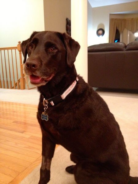
CHARLEY’S OSTEOSARCOMA HISTORY, DIAGNOSIS AND TREATMENT


Charley started limping at about 20 months old and the limping was on and off for a few days.  We took Charley to the vet after my husband was playing with him and he hugged him around his front legs and he yelped in pain.  We requested x-rays immediately (instead of the recommended course of anti-inflammatory meds and a re-check in 2 weeks).   X-rays revealed a lytic lesion in his left mid-humerus that looked like osteosarcoma except that it was not in the typical location of “away from the elbow and towards the knee”.


We were referred to Dr. Buss, an oncologist, and he did a FNA (fine needle aspirate) of the lesion and it was not cancer but rather an aneurysmal bone cyst, which is extremely rare (there are only a few documented cases).  We went back to Dr. Buss every 1-2 months to monitor the aneurysmal cyst, which was resolving on it’s own, when 7 months later Dr. Buss noticed a lytic lesion in his left proximal humerus (same bone at the cyst, but up towards his shoulder) on the x-ray.  Dr. Buss put Charley on antiobiotics for 3 weeks in case it was a bone infection (his humerus bone was already compromised because of the aneursymal cyst and an infection was a possibility) and x-rays showed improvement after week one, but worsening of the lesion after week 3.  Dr. Buss did a bone biopsy of his left proximal humerus because Charley’s history was so atypical.  The histopathology results from Charley’s bone biopsy stated:  “primary malignant neoplasia of bone; most consistent with Poorly Productive Osteogenic Osteosarcoma”.…and so began our unwanted OS journey.


Charley was 2-1/2 years old when he was diagnosed with OSA on 10/19/10.  He had his left front leg and left scapula amputated on 10/28/10 followed by 5 rounds of i.v. Carboplatin chemo every 3 weeks apart.  Charley started chemo on day 13 after his amputation, immediately after his staples were removed.  Charley’s blood work after chemo was always fine and he tolerated the Carboplatin chemo well without any side effects.  Charley had follow-up appointments with his oncologist and chest x-rays to check for lung mets every 3 months for the first year after his amputation.  After the first year post-amputation, Charley’s follow-up appointments with his oncologist and chest x-rays moved to every 6 months.


Unfortunately, we found out on 10/24/13 that Charley’s OS came back as a met underneath his amputation scar…. almost exactly 3 years to the day after his amputation.  Charley had surgery on 11/8/13 to remove the cancerous mass (and prescapular lymph node) and the surgeon was able to get clear margins around the cancerous mass (1cm at the narrowest margin because the tumor extended down to his Brachial Plexus; and 2cm at the widest margin).


The histopathology report from Charley’s cancerous mass  removal stated:  “The mass of the left prescapular incision site most likely represents the recurrance of the the prior primary osseous sarcoma.  The recurrent mass may represent telengiectatic variant of osteosarcoma.  However, morphologically is somewhat more suggestive of hemangiosarcoma suggesting that the prior mass may have indeed been hemangiosarcoma of bone origin.  Margins in relation to the mass were clean in examined sections.  I am also suspicious of local metastasis to the subscapular sinus region of the prescapular lymph node.”


Dr. Buss does not think the tumor was a hemangiosarcoma, nor was his original leg tumor.  He believes the tumor that was removed was an osteosarcoma metastasis and specifically, Telangiectatic OS, from his primary OS tumor that was in his proximal humerus (removed in October 2010).


Here’s your oncology lesson of the day:

Subclasses of osteosarcomas are determined based on the characterization of the cells as well as the type and amount of matrix present.  Subclasses include osteoblastic, chondroblastic, fibroblastic, poorly differentiated, and telangiectatic osteosarcomas; however, there is no evidence of different biological behavior between the subclasses.


Telangiectatic osteosarcoma is an unusual variant of osteosarcoma, forming 3% to 10% of all osteosarcomas.  Radiographically, these tumors appear as purely lytic destructive lesions located in the metaphyses of long bones.  The location and x-ray appearance of telangiectatic osteosarcomas are reminiscent of an aneurysmal bone cyst and can test the acumen of a diagnostic radiologist. Distinguishing between the two entities microscopically can also be quite challenging. Telangiectatic osteosarcoma shows dilated blood-filled spaces lined or traversed by septa containing atypical stromal cells, with or without production of a lacelike osteoid matrix.


Because Charley had Carboplatin after his amputation, Carboplatin can not be used again because it will not work anymore because some cancer cells survived after that chemo initially and were dormant before becoming metastatic OS.  Dr. Buss explained that he would attack this metastatic OS with 2 different chemo agents for 2 reasons:  1)  to attack the cancer from 2 different angles; and 2) to minimize the side effects of each chemo.


Charley’s chemo was  alternated between i.v. Doxorubicin (Adriamycin) and oral Lomustine (CCNU) every 3 weeks for a total of 6 rounds (Doxorubicin, Lomustine, Doxorubicin, Lomustine, Doxorubicin, Lomustine).  Charley has had 5 of 6 rounds of chemo (11/27/13: Doxorubicin; 12/18/13: Lomustine; 1/9/14: Doxorubicin; 1/29/14: Lomustine; 2/19/14: Doxorubicin).  His next and FINAL chemo #6 is Lomustine and is scheduled for 3/12/14.


Charley did take Cerenia (anti-nausea) starting on day 2 after the Doxorubicin for 4 days since the Doxo is harder on the GI system and GI side effects are the worst from days 3-5.   Lomustine is an oral chemo that is also referred to as CCNU.  It is commonly used to treat some cancers of the brain (it can cross the blood-brain barrier), lymphoma, mast cell tumors, and non-resectable soft tissue sarcomas.  Because Lomustine has a greater impact on myelosuppression (a condition in which bone marrow activity is decreased, resulting in fewer red blood cells, white blood cells, and platelets.), Charley starts antibiotics (cefpodoxine 200mg) on day 5 after chemo for 7 days to prevent an infection because of low white blood cell count (leukopenia).   Charley had his blood work done between 7-10 days (at nadir) after his first 4 rounds of chemo as well as a liver profile after his first 2 rounds of Lomustine (since Lomustine can impact liver function) and all has been fine.  Charley does not need to have blood work after his last 2 chemo rounds per Dr. Buss.


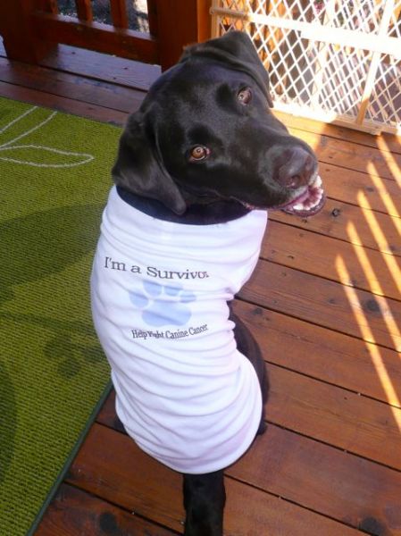
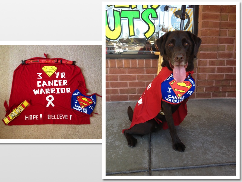
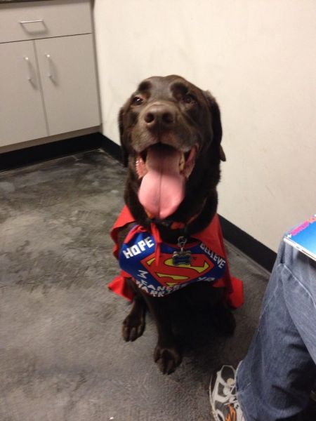
Charley is now 5-3/4 years old and he will celebrate his 40 Month Ampuversary on 2-28-14 and his 6th Birthday on 3/29/14…so he has lived over 1/2 of his life as a Tripawd and with OS, so miracles can and do happen!


CHARLEY’S PROTOCOL (3+ Year OS Warrior)


My goal with Charley’s protocol is to accomplish 2 things:  1) to support and strengthen his immune system in order to fight the cancer; and 2) to kill any rogue cancer cells by apoptosis; which will in turn give him a better quality of life and hopefully a much better quantity of life!  🙂


Here is my disclaimer about Charley’s protocol:
Every dog is different and no two cancers act exactly the same (even when comparing OS to OS, lymphoma to lymphoma, etc.).  I still tweak Charley’s protocol and nothing is written in stone.  Some pups tolerate lots of supplements without any issues and others don’t….too many supplements are not always a good thing in my opinion.  You have to do what works for you, your dog (or kitty), your family, your finances, etc.  Most importantly is to always remember to NEVER GIVE UP HOPE!!!


Since Charley’s OS returned this past October, I’ve added in Immunity4Pets and I’m back to giving the artemisinin/Artemix/Butyrex or artemisinin/artemether/Butyrex on a daily basis.  I’ve also moved his vitamin C and vitamin E from breakfast to lunch because you can’t give vitamin C within 2 hours of the Immunity4Pets.  I’ve also doubled his K9 Immunity Plus Chews to 4 chews per day.


Charley’s Protocol (68 pound, 5-3/4-year-old male neutered Lab):


Charley eats Orijen Six Fish kibble 3x day and we give Charley bottled water, not tap water because of the fluoride.


Breakfast: 7:00am
-20mg generic pepcid about 20 minutes before breakfast
-Orijen Six Fish Kibble 3/4c

-Immunity4Pets: 4 tsp sprinkled on food; dosing on container by weight (immunity4pets.com)
-Berte’s Green Blend (b-naturals.com); dosing on container by weight
-(2) K9 Immunity Plus Chews (iHerb.com)


Lunch: between 12-2pm
-Orijen Six Fish Kibble 1/2c
–Berte’s Ultra Probiotics (b-naturals.com); dosing on container by weight

-500mg of Vitamin C (Esther C)*
-400iu of Vitamin E*
-(2) 1000mg of fish oil
-(2) K9 Immunity Plus Chews

*Vitamin C and E are given daily to help flush the system so you can give the artemisinin/Artemix on a daily basis.


Dinner: between 5:30-6:00pm
-20mg generic pepcid about 20 minutes before dinner
-Orijen Six Fish Kibble 1/2c
-(2) 1000mg of fish oil


4-5 hours after dinner (between 10:30-11:00pm)
-Charley gets artemisinin (Holleypharma.com) daily with or without Artemix/artemether (Hepalin.com) wrapped in Philly Cream Cheese
-200-300mg of artemisinin along with 4 Butyrex* (pureformulas.com); when I add in Artemix or artemether I give him 1-2 caps for a total of 40-80mg of artemether


*Butyrex enhances the effect of the artemisinin/Artemix.


**Charley does not tolerate the fish oil at the 6500mg range and he gets the runs (based on the recommendations for cancer of 1000mg per 10 pounds), so he gets 4000mg/day.


I am one of the moderators on the artemisinin_and_cancer Yahoo group.  If you want more info about dosing the artes (including Artemix/artemther which is toxic at high doses), please consider joining the arte group.


Thank you for all of your prayers, positive thoughts, hugs, and kisses.  It is greatly appreciated and we can’t thank you enough for all of your support!


  Hugs from me and chocolate Labby kisses from Charley!  xoxo 


Charley survivor1

541748_10200669725242290_1775351738_n

We had Charley’s appointments this afternoon with Dr. Luther (surgeon) and Dr. Buss (oncologist).


Charley’s staples were removed…Yay!!!   Dr. Luther was excited that she got clear margins and after explaining to us how she had to cut the tumor out by feel and by using the CT imaging, it is amazing that she got clear margins especially since the tumor was around his Brachial Plexus (the nerves and arteries for his missing front leg that come off his spinal cord).  Dr. Luther gave us a tour of the facility which included the ICU, chemo room, Ultrasound, x-ray, and surgical suites (just got to peek through the outer windows).  It was very cool to get to see all the “behind the scenes”.  I asked if I could see Charley’s CT scan, so Dr. Luther brought Joe, Charley and I to her office and she went through the entire scan with us level by level which was really cool!   I gave Dr. Luther a big hug when I was leaving and thanked her for doing such a great job with Charley (twice)!


Now, on to the oncology visit…..


Dr. Buss does not think the tumor was a hemangiosarcoma, nor was his original leg tumor.  He believes the tumor that was removed was an osteosarcoma metastasis…..specifically, Telangiectatic OS…..from his primary OS tumor that was in his proximal humerus (removed in October 2010).


There was an area near the bottom of Charley’s incision site that had scabbed over.  Once the scab was removed to get to the staples, the incision was open.  Since the incision was open, Charley was not able to start chemotherapy today.  He will have his 1st chemotherapy next Wednesday 11/27, which will be i.v. Doxorubicin.  His next chemo will be 3 weeks later and it will be the oral chemo Lomustine.   Charley will alternate between the 2 chemos every 3 weeks for a total of 6 rounds (Doxorubicin, Lomustine, Doxorubicin, Lomustine, Doxorubicin, Lomustine).


Dr. Buss said that we can continue giving Charley the Immunity4Pets and Artemisinin/Artemix during chemo, so we will continue with his holistic regimen too.


Here’s your oncology lesson of the day:


Subclasses of osteosarcomas are determined based on the characterization of the cells as well as the type and amount of matrix present.  Subclasses include osteoblastic, chondroblastic, fibroblastic, poorly differentiated, and telangiectatic osteosarcomas; however, there is no evidence of different biological behavior between the subclasses.


Telangiectatic osteosarcoma is an unusual variant of osteosarcoma, forming 3% to 10% of all osteosarcomas.  Radiographically, these tumors appear as purely lytic destructive lesions located in the metaphyses of long bones.  The location and x-ray appearance of telangiectatic osteosarcomas are reminiscent of an aneurysmal bone cyst and can test the acumen of a diagnostic radiologist. Distinguishing between the two entities microscopically can also be quite challenging. Telangiectatic osteosarcoma shows dilated blood-filled spaces lined or traversed by septa containing atypical stromal cells, with or without production of a lacelike osteoid matrix.


Charley is and will always be our hero….our Super Dog, our Cancer Warrior, our miracle boy.  He has defied the odds for over 3 years and he has no plans to quit now!  We will continue our fight traversing down the familiar chemotherapy path again!


We will pray for a miracle and hope for the best!


We will pray that Charley’s journey here is far from being over, that he has many more people to inspire and bring hope to, and that he has plenty of life lessons still to teach us.


We’re not finished fighting and either is Charley.  We will continue this battle with Charley as long as he wants to fight!


Thank you for all of your prayers, positive thoughts, good karma, hugs, kisses, etc.  It is greatly appreciated and we can’t thank you enough for all of your support!  We couldn’t go down this road alone….and again….without all of you!


And most importantly, Charley is back to his Happy Labby self….and boy did we miss that smiling face!  He’s back to counter surfing, stealing food, barking loudly, jumping on the furniture, bringing us his babies, and he’s even started to do his “zoomies” again!


♥  Hugs from me and chocolate Labby kisses from Charley!  xoxo 


Charley cancer costume

Charley survivor1


Hi everyone!


I called Charley’s surgeon this afternoon to inquire about the histopathology from his mass removal and lymph node removal.  The results were in, but she did not call to tell us the results since the oncologist will be the person to discuss treatment options.  She didn’t want to go into any more detail than was given in the report since we meet with his oncologist tomorrow afternoon.  I’m glad that I called today so I could read over the report before our appointment tomorrow and have time to digest the information.


Here are Charley’s histopathology results….skip this section if you don’t want to read medical jargon!!!

__________________


DESCRIPTION:

1.  5 cm mass removed at incision (amputation) scar:

Representative sections are examined on 4 slides.  There is a somewhat multifocal and variable coalescing infiltrative neoplasm identified within the deep subcutaneous tissue.  Some areas of the neoplasm is densely cellular, other areas loosely dispersed.  Neoplastic cells are poorly differentiated and irregularly spindle-shaped with indistinct cell margins.  In some areas the cells form relatively solid haphazard sheets, sometimes with interspersed blood-filled cleft-like spaces.  In other ares the neoplastic cells form larger vascular type spaces containing variable numbers of erythrocytes.  There is little interspersed stroma.  In a few areas a small amount of collagenous-type stroma is observed.  There are also some areas where streams of eosinophillic amorphous stromal material reminiscent of ostoid are observed  within the neoplastic infiltrate.  The mitotic rate is up to 3 per high power field and the mitotic index is 22.  Mutifocal generally small areas of necrosis and hemorrhage are observed within the neoplasm.  There are some areas where small aggregrates and lobules of well-differentiated adipocyctes are identified intimately interspersed amoingst neoplastic cells/tissues.  The narrowest clean deep margin is approximately 2 cm in relation to the neoplasm.  The narrowest clean lateral margin identified is approximately 1 cm in relation to the neoplasm.


2.  Left prescapular lymph node, 1 x 3 cm tissue specimen with node

The section of lymph node with surrounding adipose tissue is examined.  Moderate hemosiderosis is observed within the lymph node.  There is also an area of vascular-like tissue proliferation within and mildly widening the subscapular sinus of the node.  Mesenchymal cells forming vascular-like spaces exhibit minimal to mild anisocytosis and anisokaryosis.  No mitotic figures are seen.


MICROSCOPIC FINDINGS:

1.  Left Prescapular Incision Site:

Poorly Differentiated Sarcoma.

Locally infiltrative.

Hemangiosarcoma versus Telangiectatic Osteosarcoma.


2.  Left Prescapular Lymph Node:

Chronic congestion with area of mildly atypical sinusoidal vascular-like proliferation.


COMMENTS:

The mass of the left prescapular incision site most likelu represents the recurrance of the the prior primary osseous sarcoma.  The recurrant mass may represent telengiectatic variant of osteosarcoma.  However, morphologically is somewhat more suggestive of hemangiosarcoma suggesting that the prior mass may have indeed been hemangiosarcoma of bone origin.  Margins in relation to the mass were clean in examined sections.  I am also suspicious of local metastasis to the subscapular sinus region of the prescapular lymph node.

__________________


In a nutshell, the good news is that the surgeon was able to get clean margins!!!  Yippee!!!!  The expected news is that were are most likely dealing with a metastasis of osteosarcoma….the unexpected news is that Charley’s original bone tumor could have been an hemangiosarcoma instead of the poorly productive osteosarcoma that was originally thought.


The bottom line is we are still dealing with a highly aggressive sarcoma as we expected.  Charley sees the surgeon tomorrow at 3pm CST  to remove his staples (he is going to be so excited because he’ll be able to move without the staples pulling) and then we see Dr. Buss, his oncologist, to discuss treatment options.  Hopefully the treatment option will be the oral chemo that he mentioned previously which is Lomustine (also known as CCNU).  I will keep everyone posted after our visits tomorrow!


Thank you for all of your prayers, positive thoughts, hugs, and kisses.  It is greatly appreciated and we can’t thank you enough for all of your support!


  Hugs from me and chocolate Labby kisses from Charley!  xoxo 


Charley collage 10.31.13

Charley collage 10.30.13


Dr. Luther, Charley’s surgeon,  just called at 12:35pm CST and Charley is out of surgery.  The breathing tube has been removed and he is on a medication drip in the ICU and appears groggy, but comfortable.


The mass was not the prescapular lymph node as they suspected, but rather a separate mass next to that node.  Going outwards, the mass attached up to the skin through the fat and muscle along part of his amputation scar.  Going inwards, the mass went all the way to his brachial plexus (the area that contains his nerves and blood vessels that supplied his missing left front leg).


Dr. Luther was able to remove the entire mass that she could visualize using the CT scan.  She was able to get 2cm margins and she took off more of the already severed (from his prior amputation) brachial plexus, but she won’t know until the biopsy comes back whether there are cancer cells still along the nerves.  She did remove the prescapular lymph node that was next to the mass because it was enlarged and that will also be sent for biopsy.


She doesn’t know if the cancer started at the skin and went deeper internally…or if it started deeper internally and progressed towards the skin, and she doesn’t know if the biopsy will be able to tell us that.  She does still feel like this is an OS met and not another type of cancer and the biopsy will hopefully answer that question also.


The good news is that Charley is recovering and the mass is out.  We are not sure yet if Charley will be able to come home today because it depends on whether his pain can be controlled without a drip and with oral medications.  If we don’t here from MVRC by 4pm CST, then we need to call to find out the plan.  If they send a picture of Charley recovering, I will post that too!


Thank you for all of your prayers, positive thoughts, hugs, and kisses.  It is greatly appreciated and we can’t thank you enough for all of your support!


  Hugs from me and chocolate Labby kisses from Charley!  xoxo 


Here’s a picture of Charley recovering in the ICU.  He’s well taken care of! 🙂


Charley 11.8.13.


Charley cancer costume

MVRC just called at 9:53am CST and Charley is under anesthesia and doing well. He’s going in now for surgery. I’ll keep praying for my boy and for another miracle.


It makes me feel so much better knowing that we are surrounded by love, hope, and prayers for Charley!


  Hugs from me and chocolate Labby kisses from Charley! xoxo 

541748_10200669725242290_1775351738_n

Charley’s surgery is tomorrow morning to remove the OS met in his lymph node that is near his amputation site.  He has to be there at 7:45am CST.  I’m not sure the exact time of his surgery, but I will keep everyone posted.  Charley can’t have anything to eat past 8:00pm CST tonight, so I fed him 2 dinners because he was not happy last Thursday when he couldn’t eat before his CT scan.  Charley thinks he hit the lotto…or his mommy is going crazy and forgot that he already ate his dinner.


We’re praying for the skill and wisdom of his surgeon, a successful surgery, clear margins (even though we are fully aware that the surgeon will may not be able to get clear margins), a quick recovery, and more precious time with our cancer warrior!


Positive thoughts, prayers, and keeping fingers and 3-paws crossed are greatly appreciated!  I will post updates tomorrow.


Here are 4 reasons that I believe are good karma for Charley’s surgery tomorrow:


1.  November is National Pet Cancer Awareness Month….maybe Charley could be their poster child!  🙂


2.  Charley won Penn Vet School’s Halloween contest on Facebook and I just found out last night!   His costume was, of course, “Superman Cancer Warrior”.  Charley’s prize is a package of Penn Vet swag.  Here’s the link to the post of Penn Vet’s FB page:

https://www.facebook.com/photo.php?fbid=10151694550011573&set=a.126537196572.115175.86740226572&type=1&theater


3. Penn Vet (University of Pennsylvania) is also where Dr. Nicola Mason is running the trial for the Bone Cancer Vaccine, so I’m thinking this is a sign!

It is now over 16 months since the first dog diagnosed with spontaneous osteosarcoma received an experimental bone cancer vaccine at the University of Pennsylvania’s School of Veterinary Medicine.  The results are highly promising and a larger phase II clinical trial is now being planned at Penn and at collaborating sites including Colorado State University and the University of Florida.  If you would like to learn more about the clinical trial, are interested in enrolling your dog, or wish to support Dr. Mason’s research, visit http://www.vet.upenn.edu/research/centers-initiatives/canine-cancer-studies.


4.  Charley received a very special present in the mail today….a beautiful handmade collar that reads “Together We Can Make a Difference” in support of Bone Cancer Dogs.  Chris O’Riley of Collars By Chris (Wellington, New Zealand) donates 50% of her profits to Bone Cancer Dogs for cancer research.  Until Chris raises $1500, she is donating 100% of her profits to Bone Cancer Dogs.


If you need a beautiful, high quality, and affordable dog collar please consider purchasing one from Collars by Chris: http://collarsbychris.weebly.com


Chris also dedicated a page for Charley on her website over a year ago!  I am still touched and honored that Charley continues to grace a page on her website.  http://collarsbychris.weebly.com/a-special-model-charley-the-hero.html


IMG_1874

IMG_1876


Thank you for all of your prayers, positive thoughts, hugs, and kisses.  It is greatly appreciated and we can’t thank you enough for all of your support!


  Hugs from me and chocolate Labby kisses from Charley! xoxo 


Here’s a few pictures of Charley modeling his new collar….and they are the “before” pictures for this surgery.  The next pictures that you’ll see after tomorrow he’ll be shaved (or at least 1/2 shaved).


IMG_1872

IMG_1869


….and my favorite collage that shows Charley’s personality!


Charley collage 10.31.13

The surgeon said the CT scan looks clear except for the cancer that’s in the prescapular lymph node.  Lungs, heart and everything else looked good.  There was some increased uptake along the border of his abdomen where she thought could be a deeper cancer or scar tissue, but she aspirated it and all she saw were fat cells.  The radiologist will have to review the scan to give a definitive, but and we won’t know until tomorrow (Friday) or Wednesday (Dr. Luther is out of the office Mon and Tues).


Charley is scheduled for surgery to remove the lymph node next Friday 11/8.  The surgeon obviously won’t be able to get clear margins because the structures near the prescapular lymph node are his jugular vein and trachea.  It would be considered a palliative surgery just like his amputation was considered (versus curative).  Dr. Luther (surgeon) has already consulted with Dr. Buss (oncologist) and the treatment options are chemo, radiation, or a combo of both based on the histopathogy of the lymph node tumor.  The surgeon says that it’s rare that OS mets to the prescapular lymph node (typically lungs or bone), but Charley’s case has not been typical since the beginning and he’s still here 3 years later.


All things considered, the news could have been worse.


Thank you for all of your prayers, positive thoughts, hugs, and kisses.  It is greatly appreciated and we can’t thank you enough for all of your support!


  Hugs from me and chocolate Labby kisses from Charley! xoxo 

Charley collage 10.31.13

Unfortunately, no surgery today for Charley. The surgeon said the cancer lump is the lymph node and that she thinks it goes into deeper structures because it’s connected at one end and that area feels hard. She said it could be scar tissue from his amp or more cancer.  A CT scan will show where the cancer is and how much is there.  I guess it’s an osteosarcoma met in a lymph node that’s not behaving typical???  Whatever it is, I just have a really bad feeling…


If they can even do surgery on Charley it wouldn’t be until end of next week. We most likely won’t have the radiology report from the CT scan until Monday.  Here’s some pictures of Charley wearing his Superman Cancer Warrior costume while we were waiting for the surgeon. Charley was just upset that he hadn’t eaten since last night!

1381165_10201905375812782_2088341282_n1441573_10201905375692779_537814426_n

MVRC just called and Charley’s awake and alert after his CT scan. He’s still in the ICU, but here’s the picture they sent. Dr. Luther, who did the MRI, will call sometime today. We get to pick Charley up this evening.

20131031_112518

Thank you for all of your prayers, positive thoughts, hugs, and kisses.  It is greatly appreciated and we can’t thank you enough for all of your support!


  Hugs from me and chocolate Labby kisses from Charley! xoxo 

Chocolate Kisses is brought to you by Tripawds.
HOME » NEWS » BLOGS » FORUMS » CHAT » YOUR PRIVACY » RANDOM BLOG