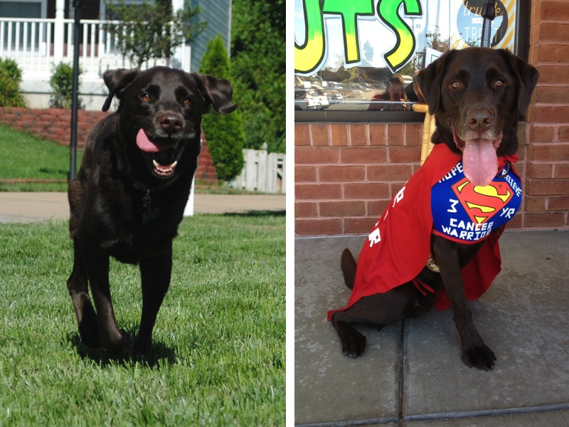
Charley 3+ Year Warrior: Osteosarcoma History, Treatment and Supplement Protocol
Posted in 3 Year Ampuversary, Amputation, Ampuversary, Artemether, Artemisinin, Artemisinin_and_cancer Yahoo group, Artemix, Berte's Green Blend, Berte's Ultra Probiotics, bloodwork, Butyrex, Cancer Survivor t-shirt, Cerenia, Chemo-Carboplatin, Chemo-Doxorubicin (Adriamycin), Chemo-Lomustine (CCNU), CT Scan, Fish Oil, Front Tripawd pics, Generic Pepcid (famotidine), Histopathology report, History/OSA diagnosis, Immunity4Pets, K9 Immunity Plus Chews, Metastatic OS, Oncology, Orijen SIx Fish, Osteosarcoma, Subclasses of OS, Supplement Protocol, Surgery to remove metastatic OS, Telangiectatic OS, Vitamin C, Vitamin E on Feb 25th, 2014
CHARLEY’S OSTEOSARCOMA HISTORY, DIAGNOSIS AND TREATMENT
Charley started limping at about 20 months old and the limping was on and off for a few days. We took Charley to the vet after my husband was playing with him and he hugged him around his front legs and he yelped in pain. We requested x-rays immediately (instead of the recommended course of anti-inflammatory meds and a re-check in 2 weeks). X-rays revealed a lytic lesion in his left mid-humerus that looked like osteosarcoma except that it was not in the typical location of “away from the elbow and towards the knee”.
We were referred to Dr. Buss, an oncologist, and he did a FNA (fine needle aspirate) of the lesion and it was not cancer but rather an aneurysmal bone cyst, which is extremely rare (there are only a few documented cases). We went back to Dr. Buss every 1-2 months to monitor the aneurysmal cyst, which was resolving on it’s own, when 7 months later Dr. Buss noticed a lytic lesion in his left proximal humerus (same bone at the cyst, but up towards his shoulder) on the x-ray. Dr. Buss put Charley on antiobiotics for 3 weeks in case it was a bone infection (his humerus bone was already compromised because of the aneursymal cyst and an infection was a possibility) and x-rays showed improvement after week one, but worsening of the lesion after week 3. Dr. Buss did a bone biopsy of his left proximal humerus because Charley’s history was so atypical. The histopathology results from Charley’s bone biopsy stated: “primary malignant neoplasia of bone; most consistent with Poorly Productive Osteogenic Osteosarcoma”.…and so began our unwanted OS journey.
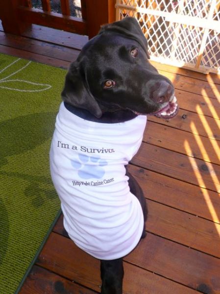
Charley was 2-1/2 years old when he was diagnosed with OSA on 10/19/10. He had his left front leg and left scapula amputated on 10/28/10 followed by 5 rounds of i.v. Carboplatin chemo every 3 weeks apart. Charley started chemo on day 13 after his amputation, immediately after his staples were removed. Charley’s blood work after chemo was always fine and he tolerated the Carboplatin chemo well without any side effects. Charley had follow-up appointments with his oncologist and chest x-rays to check for lung mets every 3 months for the first year after his amputation. After the first year post-amputation, Charley’s follow-up appointments with his oncologist and chest x-rays moved to every 6 months.
Unfortunately, we found out on 10/24/13 that Charley’s OS came back as a met underneath his amputation scar…. almost exactly 3 years to the day after his amputation. Charley had surgery on 11/8/13 to remove the cancerous mass (and prescapular lymph node) and the surgeon was able to get clear margins around the cancerous mass (1cm at the narrowest margin because the tumor extended down to his Brachial Plexus; and 2cm at the widest margin).
The histopathology report from Charley’s cancerous mass removal stated: “The mass of the left prescapular incision site most likely represents the recurrance of the the prior primary osseous sarcoma. The recurrent mass may represent telengiectatic variant of osteosarcoma. However, morphologically is somewhat more suggestive of hemangiosarcoma suggesting that the prior mass may have indeed been hemangiosarcoma of bone origin. Margins in relation to the mass were clean in examined sections. I am also suspicious of local metastasis to the subscapular sinus region of the prescapular lymph node.”
Dr. Buss does not think the tumor was a hemangiosarcoma, nor was his original leg tumor. He believes the tumor that was removed was an osteosarcoma metastasis and specifically, Telangiectatic OS, from his primary OS tumor that was in his proximal humerus (removed in October 2010).
Here’s your oncology lesson of the day:
Subclasses of osteosarcomas are determined based on the characterization of the cells as well as the type and amount of matrix present. Subclasses include osteoblastic, chondroblastic, fibroblastic, poorly differentiated, and telangiectatic osteosarcomas; however, there is no evidence of different biological behavior between the subclasses.
Telangiectatic osteosarcoma is an unusual variant of osteosarcoma, forming 3% to 10% of all osteosarcomas. Radiographically, these tumors appear as purely lytic destructive lesions located in the metaphyses of long bones. The location and x-ray appearance of telangiectatic osteosarcomas are reminiscent of an aneurysmal bone cyst and can test the acumen of a diagnostic radiologist. Distinguishing between the two entities microscopically can also be quite challenging. Telangiectatic osteosarcoma shows dilated blood-filled spaces lined or traversed by septa containing atypical stromal cells, with or without production of a lacelike osteoid matrix.
Because Charley had Carboplatin after his amputation, Carboplatin can not be used again because it will not work anymore because some cancer cells survived after that chemo initially and were dormant before becoming metastatic OS. Dr. Buss explained that he would attack this metastatic OS with 2 different chemo agents for 2 reasons: 1) to attack the cancer from 2 different angles; and 2) to minimize the side effects of each chemo.
Charley’s chemo was alternated between i.v. Doxorubicin (Adriamycin) and oral Lomustine (CCNU) every 3 weeks for a total of 6 rounds (Doxorubicin, Lomustine, Doxorubicin, Lomustine, Doxorubicin, Lomustine). Charley has had 5 of 6 rounds of chemo (11/27/13: Doxorubicin; 12/18/13: Lomustine; 1/9/14: Doxorubicin; 1/29/14: Lomustine; 2/19/14: Doxorubicin). His next and FINAL chemo #6 is Lomustine and is scheduled for 3/12/14.
Charley did take Cerenia (anti-nausea) starting on day 2 after the Doxorubicin for 4 days since the Doxo is harder on the GI system and GI side effects are the worst from days 3-5. Lomustine is an oral chemo that is also referred to as CCNU. It is commonly used to treat some cancers of the brain (it can cross the blood-brain barrier), lymphoma, mast cell tumors, and non-resectable soft tissue sarcomas. Because Lomustine has a greater impact on myelosuppression (a condition in which bone marrow activity is decreased, resulting in fewer red blood cells, white blood cells, and platelets.), Charley starts antibiotics (cefpodoxine 200mg) on day 5 after chemo for 7 days to prevent an infection because of low white blood cell count (leukopenia). Charley had his blood work done between 7-10 days (at nadir) after his first 4 rounds of chemo as well as a liver profile after his first 2 rounds of Lomustine (since Lomustine can impact liver function) and all has been fine. Charley does not need to have blood work after his last 2 chemo rounds per Dr. Buss.
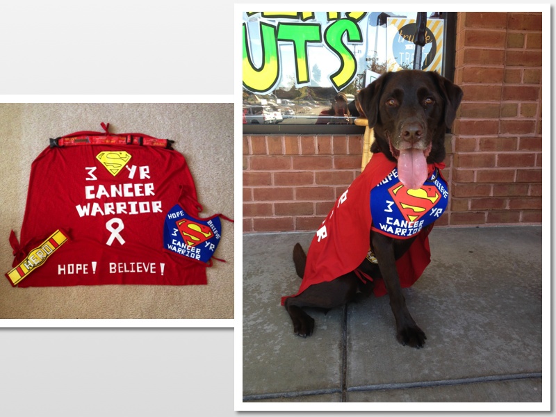
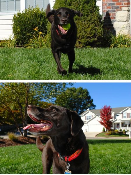
Charley is now 5-3/4 years old and he will celebrate his 40 Month Ampuversary on 2-28-14 and his 6th Birthday on 3/29/14…so he has lived over 1/2 of his life as a Tripawd and with OS, so miracles can and do happen!
CHARLEY’S PROTOCOL (3+ Year OS Warrior)
My goal with Charley’s protocol is to accomplish 2 things: 1) to support and strengthen his immune system in order to fight the cancer; and 2) to kill any rogue cancer cells by apoptosis; which will in turn give him a better quality of life and hopefully a much better quantity of life! 🙂
Here is my disclaimer about Charley’s protocol:
Every dog is different and no two cancers act exactly the same (even when comparing OS to OS, lymphoma to lymphoma, etc.). I still tweak Charley’s protocol and nothing is written in stone. Some pups tolerate lots of supplements without any issues and others don’t….too many supplements are not always a good thing in my opinion. You have to do what works for you, your dog (or kitty), your family, your finances, etc. Most importantly is to always remember to NEVER GIVE UP HOPE!!!
Since Charley’s OS returned this past October, I’ve added in Immunity4Pets and I’m back to giving the artemisinin/Artemix/Butyrex or artemisinin/artemether/Butyrex on a daily basis. I’ve also moved his vitamin C and vitamin E from breakfast to lunch because you can’t give vitamin C within 2 hours of the Immunity4Pets. I’ve also doubled his K9 Immunity Plus Chews to 4 chews per day.
Charley’s Protocol (68 pound, 5-3/4-year-old male neutered Lab):
Charley eats Orijen Six Fish kibble 3x day and we give Charley bottled water, not tap water because of the fluoride.
Breakfast: 7:00am
-20mg generic pepcid about 20 minutes before breakfast
-Orijen Six Fish Kibble 3/4c
-Immunity4Pets: 4 tsp sprinkled on food; dosing on container by weight (immunity4pets.com)
-Berte’s Green Blend (b-naturals.com); dosing on container by weight
-(2) K9 Immunity Plus Chews (iHerb.com)
Lunch: between 12-2pm
-Orijen Six Fish Kibble 1/2c
–Berte’s Ultra Probiotics (b-naturals.com); dosing on container by weight
-500mg of Vitamin C (Esther C)*
-400iu of Vitamin E*
-(2) 1000mg of fish oil
-(2) K9 Immunity Plus Chews
*Vitamin C and E are given daily to help flush the system so you can give the artemisinin/Artemix on a daily basis.
Dinner: between 5:30-6:00pm
-20mg generic pepcid about 20 minutes before dinner
-Orijen Six Fish Kibble 1/2c
-(2) 1000mg of fish oil
4-5 hours after dinner (between 10:30-11:00pm)
-Charley gets artemisinin (Holleypharma.com) daily with or without Artemix/artemether (Hepalin.com) wrapped in Philly Cream Cheese
-200-300mg of artemisinin along with 4 Butyrex* (pureformulas.com); when I add in Artemix or artemether I give him 1-2 caps for a total of 40-80mg of artemether
*Butyrex enhances the effect of the artemisinin/Artemix.
**Charley does not tolerate the fish oil at the 6500mg range and he gets the runs (based on the recommendations for cancer of 1000mg per 10 pounds), so he gets 4000mg/day.
I am one of the moderators on the artemisinin_and_cancer Yahoo group. If you want more info about dosing the artes (including Artemix/artemther which is toxic at high doses), please consider joining the arte group.
Thank you for all of your prayers, positive thoughts, hugs, and kisses. It is greatly appreciated and we can’t thank you enough for all of your support!
♥ Hugs from me and chocolate Labby kisses from Charley! xoxo ♥
