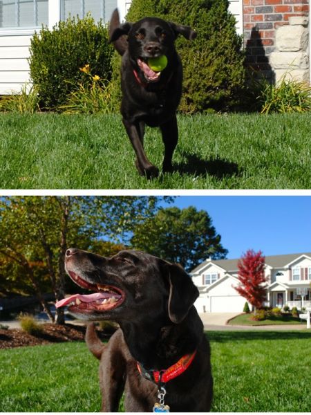
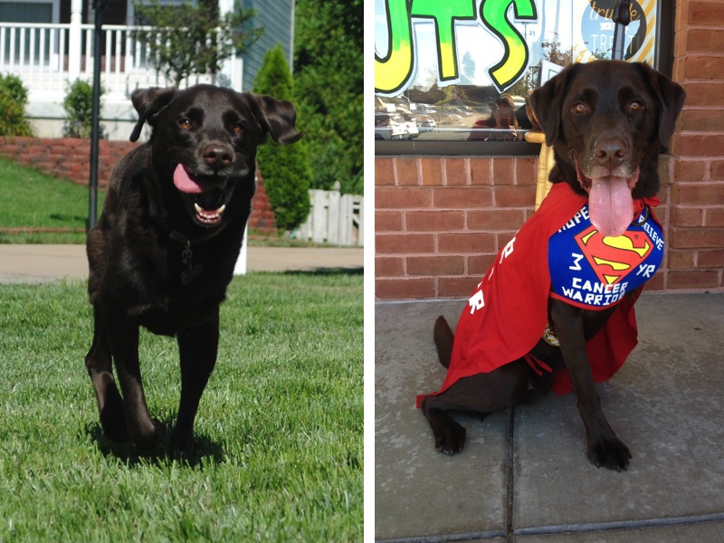
Hi everyone!
I called Charley’s surgeon this afternoon to inquire about the histopathology from his mass removal and lymph node removal. The results were in, but she did not call to tell us the results since the oncologist will be the person to discuss treatment options. She didn’t want to go into any more detail than was given in the report since we meet with his oncologist tomorrow afternoon. I’m glad that I called today so I could read over the report before our appointment tomorrow and have time to digest the information.
Here are Charley’s histopathology results….skip this section if you don’t want to read medical jargon!!!
__________________
DESCRIPTION:
1. 5 cm mass removed at incision (amputation) scar:
Representative sections are examined on 4 slides. There is a somewhat multifocal and variable coalescing infiltrative neoplasm identified within the deep subcutaneous tissue. Some areas of the neoplasm is densely cellular, other areas loosely dispersed. Neoplastic cells are poorly differentiated and irregularly spindle-shaped with indistinct cell margins. In some areas the cells form relatively solid haphazard sheets, sometimes with interspersed blood-filled cleft-like spaces. In other ares the neoplastic cells form larger vascular type spaces containing variable numbers of erythrocytes. There is little interspersed stroma. In a few areas a small amount of collagenous-type stroma is observed. There are also some areas where streams of eosinophillic amorphous stromal material reminiscent of ostoid are observed within the neoplastic infiltrate. The mitotic rate is up to 3 per high power field and the mitotic index is 22. Mutifocal generally small areas of necrosis and hemorrhage are observed within the neoplasm. There are some areas where small aggregrates and lobules of well-differentiated adipocyctes are identified intimately interspersed amoingst neoplastic cells/tissues. The narrowest clean deep margin is approximately 2 cm in relation to the neoplasm. The narrowest clean lateral margin identified is approximately 1 cm in relation to the neoplasm.
2. Left prescapular lymph node, 1 x 3 cm tissue specimen with node
The section of lymph node with surrounding adipose tissue is examined. Moderate hemosiderosis is observed within the lymph node. There is also an area of vascular-like tissue proliferation within and mildly widening the subscapular sinus of the node. Mesenchymal cells forming vascular-like spaces exhibit minimal to mild anisocytosis and anisokaryosis. No mitotic figures are seen.
MICROSCOPIC FINDINGS:
1. Left Prescapular Incision Site:
Poorly Differentiated Sarcoma.
Locally infiltrative.
Hemangiosarcoma versus Telangiectatic Osteosarcoma.
2. Left Prescapular Lymph Node:
Chronic congestion with area of mildly atypical sinusoidal vascular-like proliferation.
COMMENTS:
The mass of the left prescapular incision site most likelu represents the recurrance of the the prior primary osseous sarcoma. The recurrant mass may represent telengiectatic variant of osteosarcoma. However, morphologically is somewhat more suggestive of hemangiosarcoma suggesting that the prior mass may have indeed been hemangiosarcoma of bone origin. Margins in relation to the mass were clean in examined sections. I am also suspicious of local metastasis to the subscapular sinus region of the prescapular lymph node.
__________________
In a nutshell, the good news is that the surgeon was able to get clean margins!!! Yippee!!!! The expected news is that were are most likely dealing with a metastasis of osteosarcoma….the unexpected news is that Charley’s original bone tumor could have been an hemangiosarcoma instead of the poorly productive osteosarcoma that was originally thought.
The bottom line is we are still dealing with a highly aggressive sarcoma as we expected. Charley sees the surgeon tomorrow at 3pm CST to remove his staples (he is going to be so excited because he’ll be able to move without the staples pulling) and then we see Dr. Buss, his oncologist, to discuss treatment options. Hopefully the treatment option will be the oral chemo that he mentioned previously which is Lomustine (also known as CCNU). I will keep everyone posted after our visits tomorrow!
Thank you for all of your prayers, positive thoughts, hugs, and kisses. It is greatly appreciated and we can’t thank you enough for all of your support!
♥ Hugs from me and chocolate Labby kisses from Charley! xoxo ♥


Well HURRAH for clean margins!!!!!!!!!!!!! and HUH for the original diagnosis!? way to throw you for a loop I guess!? But at this point?…three years is three years right!? Here’s hoping the oncologist has some good suggestions and some good hope for you guys tomorrow! Looking forward to your update! WAY TO GO CHARLEY!
xoxo,
Erica & Jill
Thank you Erica and Jill!!!
Hugs and chocolate Labby kisses,
Ellen and Charley xoxo
Ellen, I’m really happy that the surgeon believes the margins were so good. I hope that, regardless of the type of cancer, Charley continues to do so well and blow raspberries at all the statistics. I hate both of these possible cancers, so I will blow raspberries with him! And just for today, let’s be grateful for the comfort getting rid of staples will bring.
Shari
Thank you Shari!!!
Hugs and chocolate Labby kisses,
Ellen and Charley xoxo
Yay for the clean margins but wow on that diagnosis now after 3 year but I agree with Shari lets all blow raspberries at it and hope Charley keeps kicking butt like he has for the past 3 years. Good luck with the oncologist tomorrow.
Hugs
Michelle & Angel Sassy
Thank you Michelle and Angel Sassy!!!
Hugs and chocolate Labby kisses,
Ellen and Charley xoxo
I know the lab results may not have been what you wanted to hear, but I also know this: Charley the SuperDog can handle whatever is thrown at him! He is awesome!! We are sending positive thoughts to Charley! Be sure to let him know how many fans he has!!!
Thank you maximutt!
Hugs and chocolate Labby kisses,
Ellen and Charley xoxo
Thinking of Charley and hopes that the followup treatment can help. Like hearing about those clean margins.
Sending healing thoughts your way.
Luanne and Spirit Shooter
Thank you Luanne and Spirit Shooter!
Hugs and chocolate Labby kisses,
Ellen and Charley xoxo
Yes! Ditto t Maxmutt! Charley is SuperDog!
Clean margkns…that’s good…..no…great!
Thanks for takng time to post this update AND pictures of that happy boy!! That Charley knows how to smile…..and how to make everyone else smile!!
If this is the “same whatever they want to call it” and he’s gone three years without “it” recurring…well, here’s to at least another three years and then some!!
Let us know now the appointment goes!
And is he completely back to being Charley (other than the stitch thing)?
Much love and hugs!
Sally and Happh Hannah
Thank you Sally and Happy Hannah!
Hugs and chocolate Labby kisses,
Ellen and Charley xoxo
Hurrah for clean margins!
The rest will be handled by Charley’s wonderful team of oncologists and his incredible pawrents.
Hope you had a great night sleep – good luck today and keep us posted.
Hugs
Linda and Tucker
Thank you Linda and Tucker!
Hugs and chocolate Labby kisses,
Ellen and Charley xoxo
Charley is our super dog! I’m glad about the clean margins but I’m sure that you were totally shocked by the suggestion that it was hemangio instead of OS three years ago. In either case, Charley is clearly an exceptional case, and I feel sure that he’ll stay that way.
Lots of Labby kisses from my Labs to Charley and your family!
Thank you KB!
Hugs and chocolate Labby kisses,
Ellen and Charley xoxo